At Which Wavelength Do You Read a Bradford Assay on a Microplate Reader
A routine analysis performed in life scientific discipline laboratories is the measurement of poly peptide concentration in liquid samples. Accurate measurement is often essential for subsequent analyses such as western blots or protein characterization.
Absorbance-based techniques are cheap, easy to handle, and well-established. They can be carried out in microplates, requiring low reagent volumes and enabling simultaneous processing of loftier sample numbers.
Spectrometer-based BMG LABTECH readers learn absorbance at discrete wavelengths or absorbance spectra between 220 and 1000 nm within a second per well, and then these assays can be easily read. This commodity discusses the most common protein quantification methods that are absorbance at 280 nm, Bradford, Bicin-Choninic Acid (BCA), and Lowry assays.
Materials and methods
- BMG LABTECH's SPECTROstar® Nano , FLUOstar® Omega, PHERAstar® FSX and CLARIOstar® plate readers with rapid and precise default settings
- Articulate 96-well plates from Greiner
- Reagents from Sigma Aldrich
- The protein standard Bovine Serum Albumin (BSA) dissolved in ddH2O to a stock of 2 mg/ml
Results and word
Absorbance at 280 nm
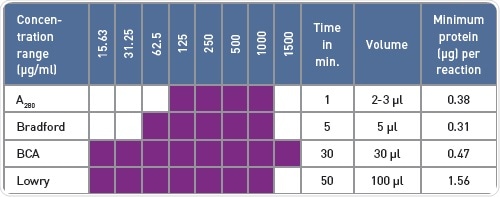
The UV light is absorbed at 280 nm wavelength by the effluvious residues of tyrosine and tryptophan amino acids (Effigy 1A), reflecting the protein concentration. This quantification method is quick and uncomplicated as information technology is not necessary to add together further reagents. The concentration range of 125-m µg/ml enabled reliable detection of BSA using the BMG LABTECH LVis plate and a book down to three µl (Figure 1B).
- three µl (BSA in ddHtwoO) on LVis plate in duplicates
- Absorbance spectrum 240-400 nm, analysis of A280
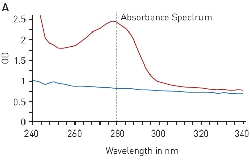
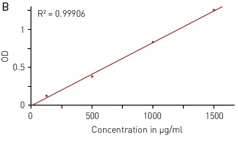
Figure 1. Total poly peptide quantitation by absorbance at 280 nm (A280) A) absorbance spectrum of water (bluish) and BSA (2 mg/ml in ddH2O, red) B) Protein standard curve of BSA. Paradigm credits: BMG Labtech.
Bradford assay
The Bradford analysis makes use of an absorbance shift of Coomassie Vivid blueish G-250 from 460 nm, in its gratis land, to 595 nm, if complexed with proteins (Figure 2A). The absorbance of poly peptide samples that are not known is related to samples containing defined protein concentrations (for instance, BSA) that are measured in parallel. A linear signal is provided by the analysis in the range of 62.v-yard µg/ml BSA (Figure 2B).
- 5 µl sample + 250 µl Bradford solution - Shake for 30 seconds
- Incubation in the dark for v to 45 minutes - Measure absorbance 400-700 nm (assay at 595 nm)
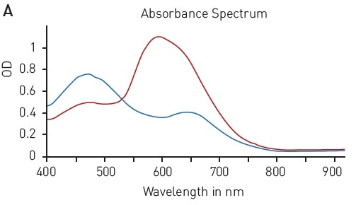
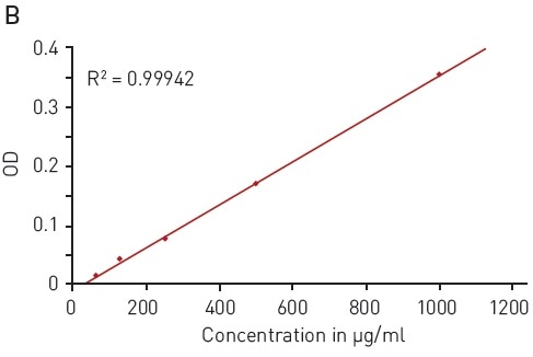
Figure ii. Bradford protein quantification assay. A) Absorbance spectrum of Coomassie Bright Bluish Thousand 250 (without protein – blue, in presence of BSA – cherry) B) Protein standard bend of BSA. Paradigm credits: BMG Labtech.
BCA assay (bicinchoninic acrid)
The ability to reduce Cu (II) sulfate to Cu+ by peptide bonds is the basis of the BCA assay. This reduction forms complexes with 2 BCA molecules, providing a chromophore with an absorbance maximum at 562 nm (Figure 3A).
In this assay, the quantification of an unknown protein sample is related to a calibration curve acquired with samples of known poly peptide concentration. With the BCA assay, a wide BSA concentration range of 15.63 to 1500 µg/ml tin can exist quantified using a hyperbola fit (Effigy 3B).
- 240 µl working reagent + 30 µl sample
- Incubate at 37 °C for xxx minutes - Measure absorbance 450-750 nm (analysis at 562 nm)
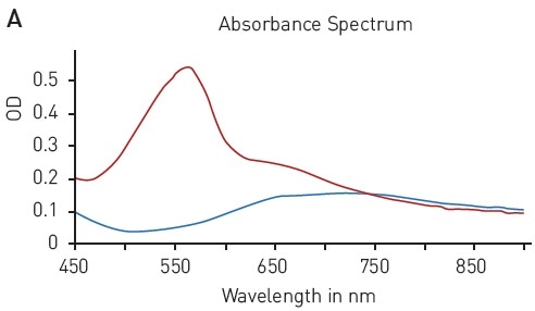
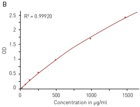
Figure iii. BCA protein quantification analysis. A) Absorbance spectrum of bicinchoninic acrid (native – bluish, BCA-Cu+-circuitous in presence of BSA - ruddy) B) Protein standard curve of BSA. Paradigm credits: BMG Labtech.
Lowry assay
Too, the modified Lowry assay is based on the ability of peptide bonds to reduce copper (Ii) sulfate. The resulting Cu+ protein circuitous reacts with Folin–Ciocalteu reagent to form a chromophore with a wide absorption spectrum of 500-800 nm (Figure 4A).
This quantification assay needs a calibration bend to be measured in parallel. With the modified Lowry assay, BSA solution tin can be reliably detected between 15.63 and grand µg/ml using a hyperbola fit (Figure 4B).
- 100 μl sample + 100 μl Lowry reagent - Incubate twenty minutes
- Add together 50 μl Folin & Ciocalteu's Phenol Regent and mix
- Incubate for 30 minutes - Mensurate 500-800 nm (analysis at 710 nm)
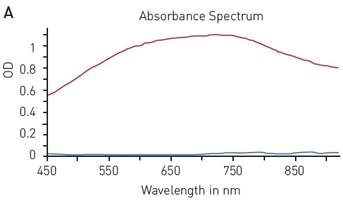
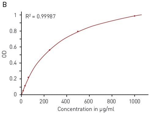
Figure four. Modified Lowry protein quantification assay. A) Absorbance spectrum of Lowry reaction (without protein - blue, in presence of BSA - red) B) Protein standard curve of BSA. Epitome credits: BMG Labtech.
Table 1. Comparison of protein quantification methods
Conclusion
BMG LABTECH readers easily detected the standard absorbance-based protein quantification assays. The protein quantification techniques differ in terms of the volume required, covered concentration range, and the time of incubation and preparation (Table ane), merely the appropriate quantification relies on other factors such every bit detergents present in the buffer that interfere differently with the assays.
Acknowledgements
Produced from materials originally authored by Andrea Krumm from BMG LABTECH GmbH, Ortenberg, Germany.
References
- Bradford, Grand.1000. (1976) Anal, Biochem., 72,248-254.
- Lowry, O.H., et al. (1951) J. Biol. Chem., 193, 265-275.
- Smith PK. et al. (1985) Anal. Biochem. ,150(1), 76-85.
About BMG Labtech

BMG LABTECH has been committed to producing microplate readers for more than than xx years. By focusing on the needs of the scientific community, the company'south innovative microplate readers have earned the company the reputation of being a engineering leader in the field.
BMG LABTECH has adult a broad range of dedicated and multi-way microplate readers for life sciences applications and high-throughput screening.
All BMG LABTECH microplate readers are "Made in Deutschland" and are conceived, developed, assembled, and tested entirely at our headquarters in Deutschland.
Since our establishment in Offenburg, Germany in 1989, BMG LABTECH has expanded to offer a worldwide sales and back up network with offices in the U.s.a., United kingdom of great britain and northern ireland, Commonwealth of australia, Japan and France. Our subsidiaries, regional offices and distributors are committed to bringing y'all innovative microplate reader technology with the quality and reliability you expect from a German company.
Our staff includes engineers and scientists from the fields of biology, biochemistry, analytical chemical science, and physics.
Sponsored Content Policy: News-Medical.cyberspace publishes articles and related content that may exist derived from sources where nosotros take existing commercial relationships, provided such content adds value to the core editorial ethos of News-Medical.Net which is to brainwash and inform site visitors interested in medical enquiry, science, medical devices and treatments.
clementandraideve1965.blogspot.com
Source: https://www.news-medical.net/whitepaper/20170403/Detection-of-Standard-Absorbance-Based-Protein-Quantification-Assays-using-BMG-LABTECH-Readers.aspx
Post a Comment for "At Which Wavelength Do You Read a Bradford Assay on a Microplate Reader"